Prostate cancer is one of the most common cancers in men. It requires accurate diagnosis, staging, and patient-centred treatment plans.
When prostate cancer is suspected, Magnetic Resonance Imaging (MRI) can be used to evaluate the presence and local extent of the cancer or suggest alternative diagnoses. By showing where the suspicious areas are in the prostate gland, MRI is used to help target ultrasound guided prostate biopsies, so the doctor doing the biopsy has the highest chance of sampling those parts of the prostate that are likely to have a tumour.
Multi-parametric MRI and Enhanced Detection of Cancerous Tissues
At TRG Imaging, we use state-of-the-art equipment, including high-field strength MRI, optimised multi-parametric pulse sequences (mpMRI), and (at some sites) artificial intelligence image reconstruction software to generate very high-resolution images of the prostate gland. This allows our specialist radiologists to identify and classify tumours, evaluate for extension beyond the prostate gland, and look for lymph node involvement and seminal vesicle invasion. Our radiologists use structured reports incorporating the ACR / ESUR PI-RADS prostate classification system to precisely communicate the results to urologists and oncologists so that each individual patient is provided with the most appropriate treatment options. Using the latest equipment and structured reporting systems also allows us to reduce the number of unnecessary biopsies by identifying very low-risk prostate nodules.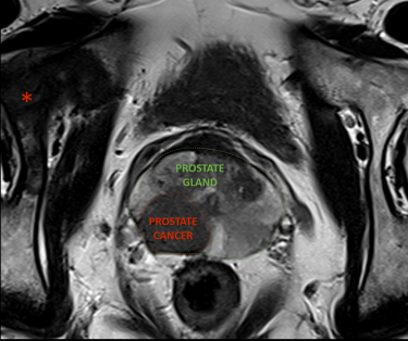
High-resolution MRI of the prostate shows two prostate cancers, one outlined in red. MRI shows that the prostate cancer has grown beyond the capsule of the prostate gland. In addition, there is a bone metastasis, shown by the asterisk.